Mass Spectrometry Imaging
Highly spatially-resolved MALDI MS imaging of biological and technical samples
Scanning Microprobe MALDI (SMALDI) mass spectrometry is a technique to analyze structured biological and technical surfaces (i.e. cell surfaces, tissue samples or intracellular compartments and synthetic structures such as biologically active surfaces) with a lateral resolution of 1-10 µm. This technique was realized in the Scanning Microprobe MALDI Time-of-Flight instrument Lamma 2000 and in the AP-SAMLDI10 ion source attached to orbital trapping mass spectrometers.
Goal of the project is the display of topological and intensity information for every detected substance as a distribution images. Further aims are an optimized sample preparation to improve sensitivity, mass resolution and effective lateral resolution.
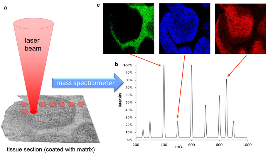
Figure: Workflow of a mass spectrometry imaging experiment (Histochem Cell Biol (2013) 139:759–783)
The tissue is sampled point by point with the laser (a). Desorbed material from each point is transfered to the mass spectrometer and a spectrum is recorded (b). Signal intensity of a component is then extracted from each mass spectrum and are transformed into gray scale values of a pixel image (c). In this way the distribution of all compound detected in the mass spectrum can be displayed.
One limiting parameter with respect to the achievable microanalytical lateral resolution is the optical resolution of the instrument (diameter of the laser spot). At the LAMMA2000 the laser spot size on the sample can be reduced to 0.7 µm at 337 nm wavelength. The AP-SMALDI10 ion source features 5 µm lateral resolution without overlapping of laser ablation areas. Commercially available instruments often offer a lower spatial resolution.
At this level of lateral resolution, the application and crystallization of matrix material is becoming the critical parameter with respect to the effective lateral resolution that can be obtained from the resulting distribution images.
In order to minimize migration of biomolecular components it is necessary to develop dedicated methods of matrix application for e.g. tissue samples. Preparation techniques such as air spraying or vapor deposition were investigated with respect to lateral migration, integration of analyte into matrix crystals and loss of effective resolution.Optimized sample preparation conditions, which lead to crystal sizes in the low micrometer range and to a homogenous distribution, were implemented in an automated fashion into the matrix sprayer SMALDIPrep.
Structured biological and artificial surfaces were investigated successfully using the new automatic data evaluation system. Biological applications demonstrate the usefulness of Scanning MALDI MS imaging in the micrometer resolution range.
Contact person
Links
