CP02
Phenotyping of pulmonary hypertension and cor pulmonale
CP02 will continue to support the CRC projects with innovative small animal imaging (echocardiography, FMT/µCT, MRI), state of the art catheterization techniques (telemetry, pressure-volume loops) and training/education of scientific and non-scientific personal in these techniques. We will perform longitudinal studies in different rat and mouse strains and genders by lung and heart imaging techniques and extensive histological analysis to correlate the observed vascular, myocardial and alveolar changes to physiological changes in the animal models of PH and RV hypertrophy/failure. Using these techniques throughout the CRC projects, we aim to establish new imaging parameters of disease severity as well as adaptive and maladaptive RV remodelling.
In recent years, imaging modalities have become indispensable tools in the basic and preclinical research. The rapidly developing high-resolution in vivo imaging technologies provide an opportunity for studying biological processes of living organisms. State of the art non-invasive small-animal imaging modalities provide qualitative and quantitative anatomical and functional information, with possibility to perform longitudinal studies allowing monitoring the disease progression. We are convinced that non-invasive imaging techniques allows us to reduce the number of animals in our research investigation according to the 3R principle (reduction, refinement, replacement). In our department we are using the most suitable imaging modalities for in vivo small-animal imaging such as ultrasonography, microscopic computed tomography (µCT) and 3D fluorescence molecular tomography (FMT) with unique approaches and methods to detect pathological processes up to microscopic level.
Overview of methods/techniques provided by Small Animal Imaging Platform
Small Animal Imaging Platform provides highly comprehensive and standardized expertise in screening, phenotyping and detailed characterization of the animal models of respiratory and cardiovascular diseases with established noninvasive imaging techniques.

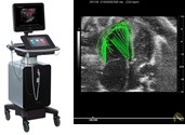
Echocardiography (Ultrasound bio-microscopy) – imaging platform is operated with Vevo3100 high-resolution ultrasound imaging system (Visaulsonics, Canada). To perform acquisition and analyses of the cardiac morphology and function in rodents from embryos to neonates and adult. Myocardial and vascular strain and strain rate analyses of the left and right ventricles, large and small arteries.
Comprehensive analyses of the segmental cardiac function, intra- and interventricular dyssynchrony in rodents with various cardiovascular pathologies. Analyses of the vascular pathologies and quantitative analyses of the vessels wall motions, vessels stiffness by PWD, vessels microanatomy and intima-media thickness (IMT).

Microscopic Computed Tomography (µCT) – Quantum GX high resolution imaging system

High resolution (up to 5 µm), high speed (8 sec), low dose x-ray imaging is suitable for longitudinal in vivo studies. Detailed analyses of the airways and lung (volume, diameter, density, aeration, functional residual capacity (FRC), tissue compartment) morphology. Analyses of the heart morphology and function (end-diastolic, end-systolic volumes, stroke volume, cardiac output, ejection fraction), including multiphase reconstruction and detailed functional analyses of the systolic and diastolic functional parameters. In vivo (lungs blood volume) and ex vivo (volume of arterial and venous systems, branches, junctions) lung and heart (coronary arteries tree) perfusion with detailed analyses of the vascular tree. µCT-derived virtual bronchoscopy useful tool for 3D visualization of the bronchial tree and diagnostic of lung pathologies including lung cancer, bronchoectases etc.
3D Fluorescence Molecular Tomography (FMT)

3D Fluorescence Molecular Tomography (FMT, PerkinElmer, USA) combined with microCT system (Quantum GX, Rigacu, Japan, PerkinElmer, USA) to investigate molecular events in whole body in response to various pathologies. Broad spectrum of near-infrared agents (spectrum 680nm, 750 nm, 800nm) available to study inflammation (MMPs and Cathepsin activities), apoptosis (annexinV), metabolism (2DG), integrin (αvβ3, αvβ5) and carbonic anhydrase IX and XII expressions, as well as vascularity, perfusion and permeability in lung, heart disease and cancer research.
The small animal imaging platform not only provides service for other departments within ILH, but also focuses on the following major objectives:
- Develop and establish a novel imaging biomarkers of pulmonary vascular and parenchymal lung diseases by in vivo and ex vivo noninvasive imaging approaches.
- Understanding the mechanisms of cardiac adaptation and maladaptation in animal models of pulmonary hypertension, lung disease and heart failure.
- Understanding the role of metabolic signaling in progression of pulmonary vascular and heart diseases.
- Develop and establish PyRadiomics approach for detailed radiological characterization of the parenchymal and vascular lung pathologies.
