Project Z3
Imaging of Systemic Bone Pathology and its Therapy using Micro- and Nano-CT Technology
3D high-resolution imaging is a pivotal technique for characterising morphology as well as mineral content of bone tissue and Biomaterials. Therefore, micro-CT is the method of choice for volume imaging when targeting 3D morphometry of microstructure and measurement of mineral contents in particular compartments of bone. Within the SFB-TRR 79, Project Z3 is dedicated to imaging and analyses of 3D micro- and nano-CT data. The spectrum of analysis encompasses:
- characterisation of animal models which are suspected to have an impaired bone status
- characterisation of bone defects in animal models aiming to mimic fractures and critical size defects in the systemically altered bone
- characterisation of biomaterials in vitro, aiming to evaluate microstructure in terms of porosity and distribution of composites
- evaluation of bone-biomaterial interactions in terms of degradation, osteo-integration as well as osteo-induction of biomaterials within bone defects
- providing 3D data for in silico analysis of bone properties.
In addition to functioning as a central service project, Z3 focuses on the development and implementation of new scanning as well as new preparation techniques for blazing new frontiers in micro-CT research while maintaining a high level of imaging quality. Furthermore, imaging and analysis of microvascularisation in bone and soft tissue are specific areas of research in the laboratory of Experimental Radiology. As Z3 is managed and implemented in the laboratory of Experimental Radiology, which is part of the Department of Diagnostic and Interventional Radiology, all state-of-the-art techniques of clinical scanning are available (MRI/US/CT/DEXA) for the purposes of the project.

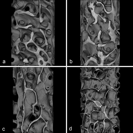
3D imaging of bone loss in iliac crest biopsies (sheep). Eight month after treatment induction, the combination of ovarectomy, deficiency diet and corticosteroid injections leads to a loss of trabecular bone (d). In comparison the control group (a) and the tretament with ovarectomy (b) and ovarectomy combined with deficiency diet (c). Cooperation : Project T1

3D-Imaging of a single Trabecle from a humeral head at a resolution of 400 nanometer isotropic voxel side length. In white the surface of the trabecle (a), in red the distribution of osteocytes (a,b)

Distribution of Microvessels in the non-fractured bone (a) vs. angiogenesis in a critical size defect (b). The 3D image post processing with an overlay of the bone contours (white) and microvessels (yellow) shows the angiogenesis of microvessels as a part of the defect healing. Cooperation : Project T2

Imaging of a Biomaterial (CPC-Cement mit 10% mesoporous bioglas). The cross-sectional image (a) shows the gray-values of cement, bioglas and air (pores). The 3D post processing with color coded differentiation between pores (green) and bioglas (white) shows the spatial distribution of these components. Cooperation : Project M2
Principal Investigators (PI) | |
|---|---|
Klinik für Diagnostische und Interventionelle Radiologie Sektionsbereich experimentelle Radiologie Contact: Klinikstraße 33 35392 Gießen Phone: +49 (0)641 / 985 41815 Fax: +49 (0)641 / 985 41809 E-Mail: marian.kampschulte@radiol.med.uni-giessen.de |
 |
Klinik für Diagnostische und Interventionelle Radiologie Sektionsbereich Bildanalyse Contact: Klinikstraße 33 35392 Gießen Phone: +49 (0)641 / 985 41836 Fax: +49 (0)641 / 985 41809 E-Mail: martin.obert@radiol.med.uni-giessen.de |
 |
Co-Workers |
|
Klinik für Diagnostische und Interventionelle Radiologie Sektionsbereich experimentelle Radiologie Contact: Schubertstraße 81 35392 Gießen Phone: +49 (0)641 / 99 39893 E-Mail: gunhild.martels@radiol.med.uni-giessen.de |
 |
