April 2013The picture shows the scanning electron microscope image of a microstructured silver electrode on a solid oxygen ion conductor. From left to right the dewetting of the silver electrode is illustrated, starting from a dense film and resulting in isolated particles, as proceeding in air after a few hours at a temperature of 400 °C. The inserts are detail pictures of micro crystallites which have been formed geometrically well-defined shapes on the surface during cooling of the sample. (Picture submitted by Jonas Neumeier.) https://www.uni-giessen.de/en/faculties/f08/departments/physchem/janek/gallerypotm/pom2013/april-2013/viewhttps://www.uni-giessen.de/@@site-logo/logo.png
Document Actions
April 2013
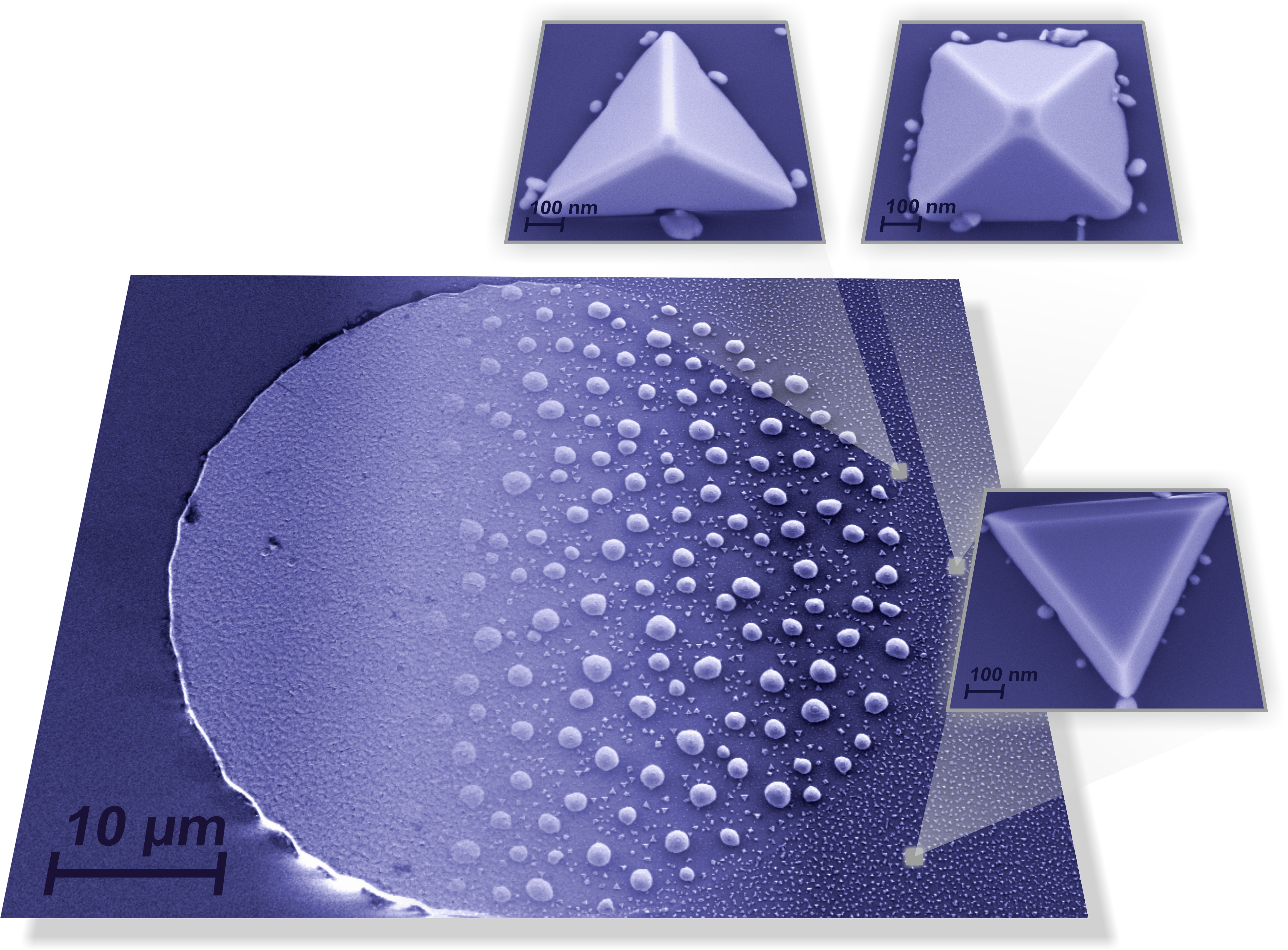
The picture shows the scanning electron microscope image of a microstructured silver electrode on a solid oxygen ion conductor. From left to right the dewetting of the silver electrode is illustrated, starting from a dense film and resulting in isolated particles, as proceeding in air after a few hours at a temperature of 400 °C. The inserts are detail pictures of micro crystallites which have been formed geometrically well-defined shapes on the surface during cooling of the sample. (Picture submitted by Jonas Neumeier.)
