July 2013The pictures show fluorescence microscopic images of human mesenchymal stem cells (hMSCs) after 2 and 24 hours of cultivation. hMSCs are stem cells from human tissue with the ability to differentiate into bone cells. On this picture, green-colored areas indicate the presence of actin (part of the cell wall), red-colored areas the presence of vinculin (component of cell-cell- and cell-matrix connections), yellow-colored areas the focal adhesions (existing cell anchorage and signaling pathways) and blue-colored areas the nucleus. After two hours of cultivation only individual cells are visible, while after 24 hours cell spreading and the formation of anchoring junctions can be seen. The aim of this imaging method is to get a first impression about the cell behavior and viability compared to control samples. Within the “Transregional Collaborative Research Centre 79” implant materials which are able to stimulate bone growth are developed and tested. The hMSCs on this picture were grown on a plasma-oxidized titanium niobium alloy. Implant samples were obtained from the Institute for Complex Materials at the IFW Dresden and plasma treated in the Janek group. All cell experiments were then carried out at the Institute of Physiological Chemistry at the TU Dresden. (Picture submitted by Markus Göttlicher.)https://www.uni-giessen.de/en/faculties/f08/departments/physchem/janek/gallerypotm/pom2013/july-2013/viewhttps://www.uni-giessen.de/@@site-logo/logo.png
Document Actions
July 2013
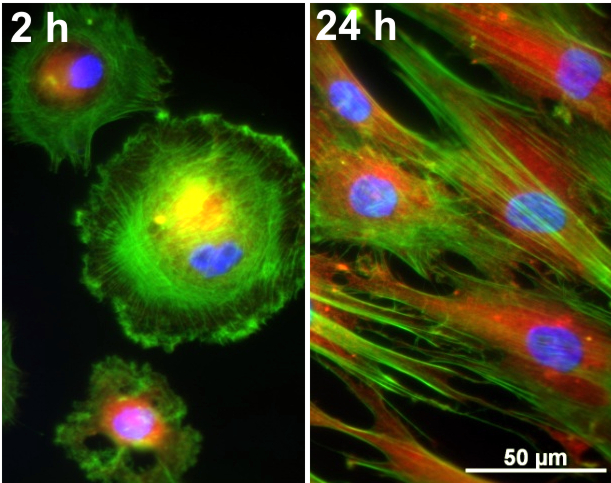
The pictures show fluorescence microscopic images of human mesenchymal stem cells (hMSCs) after 2 and 24 hours of cultivation. hMSCs are stem cells from human tissue with the ability to differentiate into bone cells. On this picture, green-colored areas indicate the presence of actin (part of the cell wall), red-colored areas the presence of vinculin (component of cell-cell- and cell-matrix connections), yellow-colored areas the focal adhesions (existing cell anchorage and signaling pathways) and blue-colored areas the nucleus. After two hours of cultivation only individual cells are visible, while after 24 hours cell spreading and the formation of anchoring junctions can be seen. The aim of this imaging method is to get a first impression about the cell behavior and viability compared to control samples. Within the “Transregional Collaborative Research Centre 79” implant materials which are able to stimulate bone growth are developed and tested. The hMSCs on this picture were grown on a plasma-oxidized titanium niobium alloy. Implant samples were obtained from the Institute for Complex Materials at the IFW Dresden and plasma treated in the Janek group. All cell experiments were then carried out at the Institute of Physiological Chemistry at the TU Dresden. (Picture submitted by Markus Göttlicher.)
