September 2013The figure shows ToF-SIMS images of different ion distributions in a human osteoblast-like cell cultured on strontium-enriched calciumphosphate cement. A 3D reconstruction of a depth profile for typical cell signals is shown in the upper image. The z axis is represented by the sputter time. An overlay of the cell signals (red) and the strontium signal (Sr+, green) can be seen in the appropriate cross-sections of the cell in the bottom images. More information can be found here (http://www.biointerphases.com/content/8/1/17/abstract). The newly generated biomaterials were made by our collaboration partner at the Technical University Dresden (group of M. Gelinsky). This project is part of the SFB Transregio 79 whose scientific aim are the development and improvement of new implant materials for the therapeutic use in systemically altered bone. (Picture submitted by Julia Kokesch-Himmelreich.)https://www.uni-giessen.de/en/faculties/f08/departments/physchem/janek/gallerypotm/pom2013/september-2013/viewhttps://www.uni-giessen.de/@@site-logo/logo.png
Document Actions
September 2013
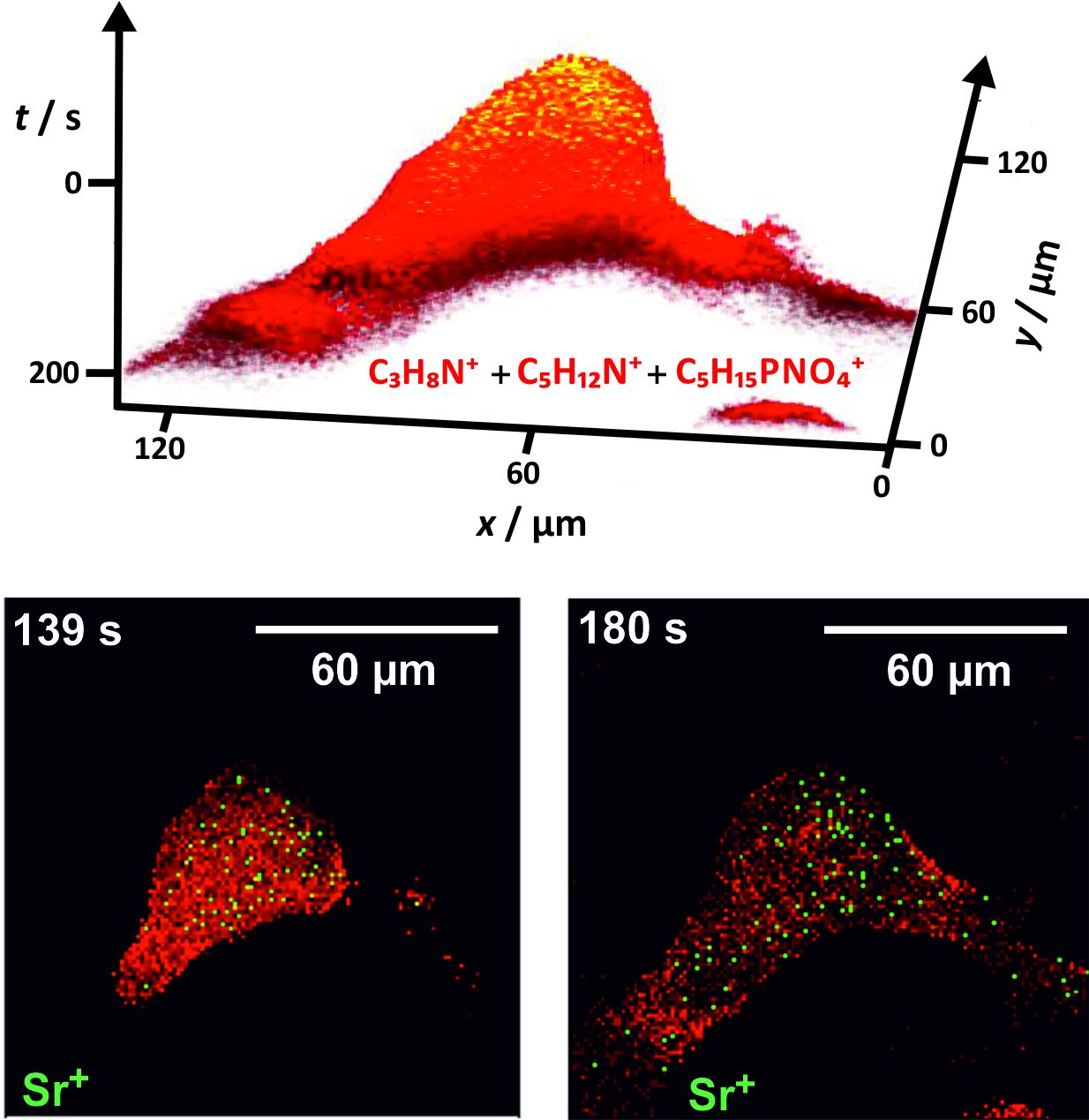
The figure shows ToF-SIMS images of different ion distributions in a human osteoblast-like cell cultured on strontium-enriched calciumphosphate cement. A 3D reconstruction of a depth profile for typical cell signals is shown in the upper image. The z axis is represented by the sputter time. An overlay of the cell signals (red) and the strontium signal (Sr+, green) can be seen in the appropriate cross-sections of the cell in the bottom images. More information can be found here (http://www.biointerphases.com/content/8/1/17/abstract). The newly generated biomaterials were made by our collaboration partner at the Technical University Dresden (group of M. Gelinsky). This project is part of the SFB Transregio 79 whose scientific aim are the development and improvement of new implant materials for the therapeutic use in systemically altered bone. (Picture submitted by Julia Kokesch-Himmelreich.)
